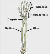
This section is a review of basic wrist anatomy. It covers the bones, ligaments, muscles and other structures that make up the wrist. For more information on how the wrist works please read the section on wrist and hand biomechanics.

What are the bones of the wrist?
The carpal bones are eight small bones that are located in the center of the wrist. These bones are divided equally into two rows. The row closest to the arm is made up of the scaphoid, lunate, triquetral and pisiform bones. The row closest to the hand is made up of the trapezium, trapezoid, capitate and hamate bones. With the palm facing upwards, the radius is the bone on the outer (lateral) aspect of the wrist and the ulna is the bone on the inner (medial) aspect of the wrist.
What is the triangular fibrocartilage complex (TFCC)?
The wrist complex also contains a “cartilage” that sits between the ulna and lateral two carpal bones. This “cartilage” is called the triangular fibrocartilage complex (TFCC). The TFCC provides stability, joint nutrition and shock absorption to the wrist.
Ligaments are like strong ropes that connect bones and provide stability to joints. In the wrist there are numerous ligaments that provide these functions. These ligaments are located on the back (dorsal), bottom (palmar), medial and lateral aspects of the wrist.
What are the bands of tissue that stabilize the wrist?
Tough bands of tissue also provide stability to the wrist. These bands are known as the extensor retinaculum and the flexor retinaculum. The extensor retinaculum is located on dorsal aspect of the wrist. The flexor retinaculum is located on the palmar aspect of the wrist. The carpal tunnel is a tunnel that contains the median nerve and tendons that flex the fingers and thumb. The “floor” and “walls” of the tunnel are formed by the carpal bones. The flexor retinaculum forms the “roof” of the carpal tunnel.
What are the attachments of the muscles and tendons that cross the wrist?
Tendons connect muscles to bone. Many of the muscles that move the wrist originate from the elbow and attach to the bones of the wrist. Many of the muscles that move the fingers and thumb also originate from the elbow. The tendons of these muscles cross the wrist and attach to the bones of the hand. The large muscles that bend (flex) the wrist, fingers and thumb originate from the medial aspect of the elbow. The large muscles that straighten (extend) the wrist, fingers and thumb originate from the lateral aspect of the elbow. Other muscles that help to turn the wrist and palm down (pronate) and turn the wrist and palm up (supinate) are located deep in the forearm.
What are the major nerves of the wrist?
Finally, the median and ulnar nerves are the major nerves of the wrist and hand. These nerves begin in the neck, run the length of the arm, through the carpal tunnel and into the hand. These nerves transmit electrical impulses to and from the brain to move and provide feeling to the hand
Visit Joint Pain Info for information on other joint injuries and problems.
A recommended book for learning wrist anatomy:

Moore’s Clinically Oriented Anatomy
Renowned for its comprehensive coverage and engaging, storytelling approach, the bestselling Moore’s Clinically Oriented Anatomy, 9th Edition, guides students from initial anatomy and foundational science courses through clinical training and practice. A popular resource for a variety of programs, this proven text serves as a complete reference, emphasizing anatomy that is important in physical diagnosis for primary care, interpretation of diagnostic imaging, and understanding the anatomical basis of emergency medicine and general surgery.
