
This section is a review of basic elbow anatomy. It covers the bones, ligaments, muscles and other structures that make up the elbow. For more information on how the elbow works please read the section on basic elbow biomechanics.
What bones make up the elbow?
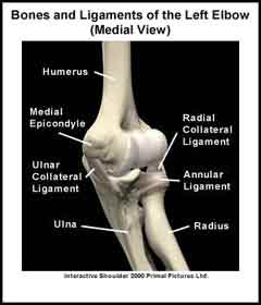
The elbow is made up of three bones, which are connected by muscles, ligaments and tendons. The humerus is the large upper arm bone. The ulna and radius are the two bones in the forearm. Looking at the forearm with the palm of the hand facing up, the ulna is located on the inner (medial) aspect of the forearm. The radius is located on the outer (lateral) aspect of the forearm. Projecting from the end of the humerus are the medial and lateral epicondyles. The epicondyles are the boney attachment sites for many of the forearm muscles.
How many joints are in the elbow?
The elbow joint is actually three separate joints; the ulnohumeral joint, the radiohumeral joint and the superior radioulnar joint. These three joints are enclosed by a loose “bag” called the joint capsule. Movement between the ulna and the humerus occurs at the ulnohumeral joint. Movement between the radius and the humerus occurs at the radiohumeral joint and movement between the radius and the ulna occurs at the superior radioulnar joint.
What are the ligaments of the elbow?
Ligaments are like strong ropes that connect bones and provide stability to joints. In the elbow there are four main ligaments. On medial aspect of the elbow is the ulnar collateral ligament that connects the ulna to the humerus. On lateral aspect of the elbow is the radial collateral ligament that connects the radius to the humerus. The other two ligaments are the annular ligament and the quadrate ligament. They connect the radius to the ulna.
Articular cartilage is a smooth shiny material that covers the ends of the bones in the elbow. There is articular cartilage anywhere that two bony surfaces come into contact with each other. In the elbow, articular cartilage covers the ends of the humerus, radius and ulna. Articular cartilage allows the elbow bones to move easily as the elbow bends (flexes), straightens (extends), rotates the palm up (supinates), and rotates the palm down (pronates).
What muscles move the elbow?
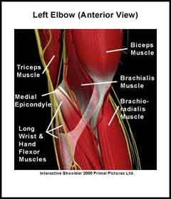
The strong biceps, brachialis and brachioradialis muscles flex the elbow. The triceps muscle extends the elbow. Other muscles that move the hand at the wrist originate at the elbow. These muscles attach via tendons to the medial and lateral epicondyles. The forearm muscles that originate on the medial epicondyle help to flex the wrist and hand. The forearm muscles that originate on the lateral epicondyle help to extend the wrist and hand. There are also muscles located deep in the elbow that help to supinate and pronate the forearm and hand.
How many bursa/bursae are there in the elbow?
Finally, a bursa (pl. bursae) is a small fluid-filled sac that decreases the friction between two tissues. Bursae also protect bony structures. There are three different bursae around the elbow the largest of which is the olecranon bursa in the back of the elbow. Normally, a bursa has very little fluid in it but if it becomes irritated it can fill with fluid.
A recommended book for learning elbow anatomy:
Moore’s Clinically Oriented Anatomy
Renowned for its comprehensive coverage and engaging, storytelling approach, the bestselling Moore’s Clinically Oriented Anatomy, 9th Edition, guides students from initial anatomy and foundational science courses through clinical training and practice. A popular resource for a variety of programs, this proven text serves as a complete reference, emphasizing anatomy that is important in physical diagnosis for primary care, interpretation of diagnostic imaging, and understanding the anatomical basis of emergency medicine and general surgery.