
In order to understand this condition, it is important to understand the anatomy and function of the foot. Please read Foot Pain Info’s section on basic ankle anatomy. For additional background information on the biomechanics of the foot please read Foot Pain Info’s section on basic foot and ankle biomechanics.
What is the tibialis posterior tendon and what is its function?
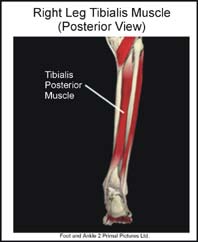
Tendons are “rope like” structures that connect muscles to bones. Many of the muscles that move the foot are found in the lower leg. These muscles attach via their tendons to various bones in the foot. The muscles that move the foot inwards (invert the foot) originate deep on the back of the lower leg. One of these muscles, the tibialis posterior, has a tendon that wraps around the inner ankle bone (medial malleolus) and attaches to the bones that form the inside arch of the foot. Accordingly the tibialis posterior muscle and its tendon are important supporting structures for the medial arch.
What is tibialis posterior tendonitis?
Tibialis posterior tendonitis is the term used to describe irritation (inflammation) of the tibialis posterior tendon. A common site for tibialis posterior tendonitis to occur is in the area behind the medial malleolus. Although this site is the most common, inflammation can occur anywhere along the tibialis posterior tendon.
What does tibialis posterior tendonitis feel like?
Tibialis posterior tendonitis usually begins with a gradual onset of dull, intermittent pain behind the medial malleolus or in the inside arch of the foot. It may progress and develop into a sharp continuous pain. Running, lunging or jumping can increase the pain. Tenderness and swelling is often found along the course of the tibialis posterior tendon.
What causes tibialis posterior tendonitis?
Tibialis posterior tendonitis usually develops as a result of overuse. Activities such as jumping, running, walking or even prolonged standing can cause undue stress on the tibialis posterior tendon. This in turn leads to the development of microscopic tears in the tibialis posterior tendon resulting in inflammation and pain. Poor flexibility of the calf muscles and of the Achilles tendon, overpronation (feet rolled in), inappropriate footwear and increasing age are some of the other factors that can cause tibialis posterior tendonitis.
Finally, tibialis posterior tendonitis can develop as a result of direct trauma, after an ankle sprain, or after an ankle fracture.
Can tibialis posterior tendonitis be detected on X-rays?
Inflammation of the tibialis posterior tendon cannot be seen on x-ray. Therefore, although x-rays are often done to rule out bony injuries in individuals with tibialis posterior tendonitis these x-rays are usually normal. Diagnostic ultrasound of the tendon can be performed to assess the integrity of the tendon. Other diagnostic tests, such as bone scans or MRI’s, are not usually required in typical cases of tibialis posterior tendonitis though blood work may be required to rule out other diseases that may be associated with tibialis posterior tendonitis such as rheumatoid arthritis.
What is the treatment for tibialis posterior tendonitis?
The treatment of tibialis posterior tendonitis should be individualized. The most important first step in the treatment (and prevention) of tibialis posterior tendonitis is to wear good, supportive shoes. Further treatment may include relative rest and icing to decrease pain around the tibialis posterior tendon, stretching and strengthening exercises, shoe orthotics or medications. A cortisone injection may be required to reduce inflammation and pain. If the above measures are not successful surgery may be necessary.
What other information is available on tibialis posterior tendonitis?
Foot Pain Info’s links section has additional information on this topic. Links have been provided to other websites as well as online medical journals. Visit Joint Pain Info for information on other joint injuries and problems.