
Biomechanics is the term used to describe movement of the body. This section is a review of basic foot and ankle biomechanics. In order to understand the biomechanics of the foot and ankle it is important to understand their anatomy. Please read the sections on basic foot anatomy and basic ankle anatomy before reading this section.
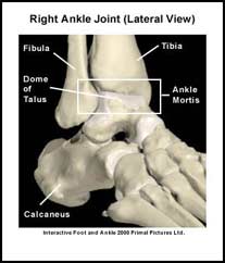
The ankle is a modified hinge joint. It plays a key role in transferring the forces from the foot to the leg. The ankle joint is made up of three bones, which are connected by ligaments, muscles and tendons. A strong ligament joins the ends of the tibia and fibula to form the ankle “mortis”. The “dome” of the talus (the highest bone of the foot) fits inside the ankle mortis to form the ankle joint. The ankle allows movement in only one plane. It allows the foot to move upwards (dorsiflexion) and downwards (plantar flexion).
The foot is made up of 26 bones. There are numerous joints between these bones that allow the foot to be both a rigid lever and a shock absorber. The largest joint in the foot is the subtalar joint. Inward movement of the foot (inversion), and outward movement of the foot (eversion) occur primarily at the subtalar joint.
The normal end ranges of motion for the foot and ankle vary between individuals and between children and adults. The following are approximate end ranges of motion for adults:
• Dorsiflexion – 20 degrees
• Plantar flexion – 60 degrees
• Eversion – 15 degrees
• Inversion – 35 degrees
The gait cycle (walk cycle) describes what happens to the foot and ankle from the point of initial contact of one foot with the ground to the point at which the same foot contacts the ground again. The gait cycle is divided into the swing phase and the stance phase. During the swing phase the foot is not in contact with the ground. As the name implies it is the phase of the gait cycle in which the foot swings forward to take another step. During the stance phase the foot is in contact with the ground. The stance phase of the gait cycle can also be divided into three stages. The first stage is called heel strike, the second stage is called mid-stance, and heel lift is the final stage. The biomechanics of the foot are best explained by describing what happens to the foot during the stance phase of the gait cycle.
During heel strike of the stance phase the foot begins to pronate. Pronation of the foot is the term that describes the rolling motion of the foot inwards and flattening of the inner (medial) arch of the foot. Pronation allows the foot to adapt to uneven terrain and absorb the impact of the foot striking the ground. It is during this phase that the foot begins to act like a shock absorber.
During midstance the entire foot is in contact with the ground and the weight of the body is directly over the foot. It is during this phase that the foot is maximally pronated. The foot acts as a shock absorber during the early part of this phase. As the body weight shifts forward the foot begins to return to a neutral position in preparation for heel lift.
Heel lift occurs at the end of the stance phase. Supination of the foot is the term used to describe the rolling motion of the foot outwards and the rising of the inner (medial) arch of the foot. During heel lift, the foot supinates to act as a rigid lever. The plantar fascia is a strong connective tissue that runs along the bottom of the foot connecting the heel to the base of the toes. The bones, muscles and the plantar fascia act together to form this rigid lever.
Abnormal amounts of pronation or supination can cause a variety of foot and leg problems. Abnormal pronation (overpronation) occurs as a result of the foot pronating when it should be neutral or supinating. Abnormal supination occurs when the foot is too rigid. These abnormal biomechanics can create lower back, hip, knee, ankle and/or foot problems.