Calcaneal Apophysitis
In order to understand this condition, it is important to understand the anatomy and function of the foot. Please read Foot Pain Info’s section on basic foot anatomy. For additional background information on the biomechanics of the foot please read Foot Pain Info’s section on basic foot and ankle biomechanics.

What is the calcaneal apophysis?
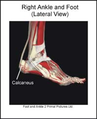
Many of the muscles that move the foot are found in the lower leg. These muscles attach via tendons to various bones in the foot. The main muscles that move the foot downwards (plantar flex the foot) and propel the body forward are the calf muscles (gastrocnemius and soleus muscles). These muscles merge to form a ‘rope like’ structure called the Achilles tendon. In growing young people the Achilles tendon attaches to the heel bone (calcaneus), at a point called the calcaneal apophysis. This is an area in the back of the calcaneus where the bone grows.
What is Sever’s disease (calcaneal apophysitis)?
Sever’s disease (calcaneal apophysitis) is the term used to describe irritation (inflammation) of the calcaneal apophysis. This condition often occurs before or during the growth spurt in boys and girls, or shortly after they begin a new activity. Sever’s disease is common is running and jumping sports.
What does Sever’s disease feel like?
Sever’s disease usually develops gradually. The pain from Sever’s disease is often intermittent and localized to the area where the Achilles tendon attaches to the calcaneus. Swelling may be noted in this area. There can be tenderness on squeezing the calcaneus or pain when trying to stretch the calf muscles. Occasionally there is night pain. As Sever’s disease progresses there can be continuous pain.
What causes Sever’s disease?
Sever’s disease usually develops as a result of overuse and is common in active children between the ages of 8 to 12. Activities that involve running or jumping can cause undue stress on the calcaneal apophysis. This in turn leads to the development of microscopic damage to the calcaneal apophysis resulting in inflammation and pain. Poor flexibility of the calf muscles and of the Achilles tendon, overpronation (feet rolled in) and inappropriate footwear are some of the other factors that can cause Sever’s disease.
Can Sever’s disease be detected on X-rays?
Low-grade inflammation of the calcaneal apophysis cannot be seen on x-ray. Therefore, although x-rays are often done to rule out bony injuries in children with Sever’s disease these x-rays are usually normal. Advanced Sever’s disease can be seen on x-ray but usually the problem is treated before it reaches this point. Other diagnostic tests, such as bone scans or MRI’s, are not usually required in typical cases of Sever’s disease. These, or other tests, may be required to rule out other conditions, such as stress fractures of the calcaneus or other bony abnormalities that can mimic Severs disease.
What is the treatment for Sever’s disease?
The treatment of Sever’s disease should be individualized. The most important first steps in the treatment of Sever’s disease are activity modification (including rest and sometimes crutches) and good shoes. Further treatment may include icing to decrease pain around the calcaneal apophysis, stretching and strengthening exercises, shoe orthotics or medications to relieve pain. Rarely, a removable cast is necessary to completely rest the foot.
What other information is available on Sever’s disease?
Foot Pain Info ‘s links section has additional information on this topic. Links have been provided to other websites as well as online medical journals. Visit Joint Pain Info for information on other joint injuries and problems.
