
In order to understand this condition, it is important to understand the anatomy and function of the foot. Please read Foot Pain Info’s section on basic foot anatomy. For additional background information on the biomechanics of the foot please read Foot Pain Info’s section on basic foot and ankle biomechanics.
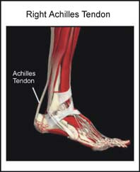
What is the Achilles Tendon and what is its function?
Many of the muscles that move the foot are found in the lower leg. These muscles attach via tendons to various bones in the foot. The main muscles that move the foot downwards (plantar flex the foot) and propel the body forward are the calf muscles (gastrocnemius and soleus muscles). These muscles are connected to the heel bone (calcaneus) by the “rope like” Achilles tendon.
What is Achilles Tendonitis?
Achilles Tendonitis is the term used to describe irritation (inflammation) of the Achilles tendon. The most common site for Achilles Tendonitis to occur is an area 2 – 6 cm. (1 – 2.5 in.) above where the tendon attaches to the calcaneus. Although this site is the most common area for Achilles Tendonitis to occur, inflammation can occur anywhere along the Achilles tendon.
What does Achilles Tendonitis feel like?
Achilles Tendonitis usually develops gradually. The pain from Achilles Tendonitis is often localized to the area 2 to 6 cm. above the Achilles tendon’s attachment to the calcaneus. Swelling is often noted in this area. There can be tenderness on squeezing the Achilles tendon or pain when trying to stretch the calf muscles. Pain and stiffness are usually worse in the morning. They are often relieved by activity only to recur after activity stops. As Achilles Tendonitis progresses there can be continuous pain.
What causes Achilles Tendonitis?
Achilles Tendonitis usually develops as a result of overuse. Activities such as jumping, running, or even walking can cause undue stress on the Achilles tendon. This in turn leads to the development of microscopic tears in the Achilles tendon resulting in inflammation and pain. Poor flexibility of the calf muscles and of the Achilles tendon, overpronation (feet rolled in), high arched feet, inappropriate footwear and increasing age are some of the other factors that can cause Achilles Tendonitis.
Can Achilles Tendonitis be detected on X-rays?
Inflammation of the Achilles tendon cannot be seen on x-ray. Therefore, although x-rays are often done to rule out bony injuries in individuals with Achilles Tendonitis these x-rays are usually normal. Sometimes the body will form calcium in areas of inflammation and occasionally a spur can be seen at the attachment of the Achilles tendon to the calcaneus. Rarely calcification can be seen within the Achilles tendon. If the Achilles tendon is swollen, diagnostic ultrasound of the tendon can be performed to assess the integrity of the tendon. Other diagnostic tests, such as bone scans or MRI’s, are not usually required in typical cases of Achilles Tendonitis though blood work may be required to rule out other diseases that may be associated with Achilles Tendonitis such as rheumatoid arthritis.
What is the treatment for Achilles Tendonitis?
The treatment of Achilles Tendonitis should be individualized. The most important first step in the treatment (and prevention) of Achilles Tendonitis is to wear proper shoes. Further treatment may include relative rest and icing to decrease pain around the Achilles tendon, stretching and strengthening exercises, shoe orthotics or medications. Ultrasound and other forms of physiotherapy may also be helpful.
What other information is available on Achilles Tendonitis?
Foot Pain Info ‘s links section has additional information on this topic. Links have been provided to other websites as well as online medical journals. Visit Joint Pain Info for information on other joint injuries and problems.