
This section is a review of basic ankle anatomy. It covers the bones, ligaments, muscles and other structures that make up the ankle. For more information on how the ankle works please read the section on basic foot and ankle biomechanics.
What type of joint is the ankle?
The ankle is a synovial hinge joint. A hinge joint allows movement in one plane, typically flexion and extension. In the case of the ankle, these movements are called plantarflexion and dorsiflexion.
What are the bones of the ankle?
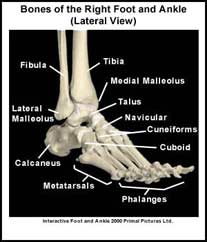
The ankle is made up of three bones; the tibia, the fibula, and the talus. The bones are connected by muscles ligaments and tendons. The tibia is the large bone located on the inner (medial) aspect of the shin. The fibula is the smaller bone located on the outer (lateral) aspect of the shin. The ends of the tibia and fibula are joined together by a strong ligament to form a socket called the ankle “mortise”. The talus is the highest bone of the foot. It has a “dome” that fits inside the ankle mortise to form the ankle joint.
The ankle links the foot to the lower leg. The bony structure on the lateral aspect of the ankle is called the lateral malleolus. It is formed by the end of the fibula. The bony structure on the medial part of the ankle is called the medial malleolus. It is formed by the end of the tibia. The medial and lateral malleoli are the bony attachment sites for the ankle ligaments.
What is the articular cartilage of the ankle?
Articular cartilage is a smooth shiny material that covers the ends of the bones in the ankle. There is articular cartilage anywhere that two bony surfaces come into contact with each other. In the ankle, articular cartilage covers the end of the tibia, the dome of the talus and a small part of the fibula. Articular cartilage allows the ankle bones to move easily as the ankle bends up (dorsiflexes), and bends down (plantarflexes).
What are the ligaments of the ankle?
Ligaments are like strong ropes that help connect bones and provide stability to joints. In the ankle, there are three ligaments on the lateral aspect of the ankle and one broad ligament on the medial aspect of the ankle. It is most common for people to injure the ligaments on the lateral aspect of the ankle.
What tendons cross the ankle?
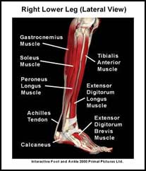
Tendons connect muscles to bone. Many of the muscles that move the foot originate from the lower leg. The tendons of these muscles cross the ankle and attach to various bones in the foot. The muscles that move the foot upwards (dorsiflex the foot) originate on the front of the lower leg. The muscles that move the foot outwards (evert the foot) originate on the lateral aspect of the lower leg. The muscles that move the foot inwards (invert the foot) originate deep on the back of the lower leg. The muscles that move the foot downwards (plantarflex the foot) and propel the body forward originate from the knee and the back of the lower leg. The muscles that play the largest role in propulsion are the calf muscles (gastrocnemius and soleus muscles). These muscles join to form the Achilles tendon that attaches onto the heel bone (calcaneus).
How many bursa/bursae are in the ankle?
Finally, a bursa (pl. bursae) is a small fluid-filled sac that decreases the friction between two tissues and protects bony structures. There are many different bursae around the ankle. The two that are commonly injured are the bursae that protect the medial and lateral malleoli. Normally, a bursa has very little fluid in it but if it becomes irritated it can fill with fluid. This is called bursitis.
A recommended book for learning ankle anatomy:
Moore’s Clinically Oriented Anatomy
Renowned for its comprehensive coverage and engaging, storytelling approach, the bestselling Moore’s Clinically Oriented Anatomy, 9th Edition, guides students from initial anatomy and foundational science courses through clinical training and practice. A popular resource for a variety of programs, this proven text serves as a complete reference, emphasizing anatomy that is important in physical diagnosis for primary care, interpretation of diagnostic imaging, and understanding the anatomical basis of emergency medicine and general surgery.