
This article is a review of the anatomy of the hip. It covers the bones, ligaments, muscles and other structures that make up the hip joint.
What type of joint is the hip?
The hip joint is a ball and socket joint. The ball is formed by the top of the thigh bone (the femur) and is called the “head” of the femur. The socket is formed by the bones of the pelvis and is called the acetabulum. Muscles, ligaments and tendons help hold the head of the femur in the acetabulum (the ball in the socket).
Articular cartilage is a smooth shiny material that covers the head of the femur and the acetabulum. Articular cartilage covers the bony surfaces wherever they come into contact with each other. Articular cartilage allows the head of the femur to move easily inside the acetabulum as the leg moves. Fluid also helps the head of the femur move easily inside the acetabulum. This fluid (called synovial fluid) provides nourishment and lubrication to the hip joint.
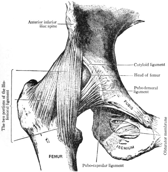
The hip joint is surrounded by a strong “bag” called a joint capsule. Ligaments are like strong ropes that help connect bones and provide stability to joints. Ligaments reinforce the capsule and connect the head of the femur to the acetabulum. These ligaments help prevent the head of the femur from coming out of the acetabulum. Larger, stronger ligaments also provide stability to the hip joint.
What is the acetabulum of the hip?
The acetabulum is the concave joint surface on the pelvis that articulates with the head of the femur. It is covered with articular cartilage and has a ring of tissue around it called the labrum. The labrum also helps provide stability to the hip.
Tendons connect muscles to bone. There are many muscles that surround the hip joint. These muscles and their tendons provide stability to the hip joint when the leg is moved. These muscles are also necessary for activities such as walking, running and jumping.
What are the muscles that control movement at the hip?
The hamstring muscles (at the back of the leg) act with the gluteus maximus (the “butt muscle”) to move the leg backwards at the hip. The hip flexors (iliopsoas and rectus femoris) move the leg forward at the hip. The groin muscles (adductor magnus and longus) move leg toward the midline of the body. The abductor group (gluteus medius, gluteus minimus and tensor fascia lata) move the leg away from the body and are also responsible for stabilizing the hip joint during weight bearing activities.
Finally, a bursa (pl. bursae) is a small sac of fluid that decreases friction between tendons, muscles and bones. The main bursa of the hip joint is the bursa of the greater trochanter. This bursa is outside of the hip joint. It can become injured by direct contact (falling directly onto the outside of the hip), or irritated from overuse. This can result in trochanteric bursitis.
Hip Pain Info’s links section has additional information on osteonecrosis of the hip. Links have been provided to other websites as well as online medical journals. Visit Joint Pain Info for information on other joint injuries and problems.
A recommended book for learning hip anatomy:
Moore’s Clinically Oriented Anatomy
Renowned for its comprehensive coverage and engaging, storytelling approach, the bestselling Moore’s Clinically Oriented Anatomy, 9th Edition, guides students from initial anatomy and foundational science courses through clinical training and practice. A popular resource for a variety of programs, this proven text serves as a complete reference, emphasizing anatomy that is important in physical diagnosis for primary care, interpretation of diagnostic imaging, and understanding the anatomical basis of emergency medicine and general surgery.