
The foot is made up of 26 bones, which are divided into three sections called the rearfoot, midfoot and forefoot. The talus and calcaneus (heel bone) are the bones that make up the rearfoot. The talus is the highest bone in the foot and it is also part of the ankle. The calcaneus is the largest bone in the foot. It sits below the talus. The navicular, cuboid and the three cuneiforms are the bones that make up the midfoot. The five metatarsals and nine phalanges are the bones that make up the forefoot.
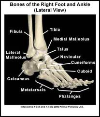
There are three arches in the foot. There is an inner (medial) arch, an outer (lateral) arch and an arch in the forefoot called the transverse arch. Ligaments are like strong ropes that connect bones and provide stability to joints. In the foot there are numerous ligaments that support the arches and stabilize the bones. These ligaments are located on the top (dorsal), bottom (plantar) medial and lateral aspects of the foot.
The plantar fascia is a key structure that helps support the medial and lateral arches of the foot. The plantar fascia is a strong connective tissue that runs along the bottom of the foot connecting the heel to the base of the toes. When weight is put on the foot the plantar fascia helps to “lock” the bones of the foot and stabilizes these arches.
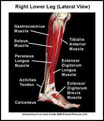
Many of the muscles that move the foot originate from the lower leg. These muscles attach via tendons to various bones in the foot. The muscles that move the foot upwards (dorsiflex the foot) originate on the front of the lower leg. The muscles that move the foot outwards (evert the foot) originate on the lateral aspect of the lower leg. The muscles that move the foot inwards (invert the foot) originate deep on the back of the lower leg. The muscles that move the foot downwards (plantarflex the foot) and propel the body forward originate from the knee and the back of the lower leg. The muscles that play the largest role in propulsion are the calf muscles (gastrocnemius and soleus muscles). These muscles join to form the Achilles tendon that attaches onto the calcaneus. In addition to the long muscles, there are also numerous short muscles in the foot. These muscles also play a role in stabilizing the arches of the foot and in moving the toes.
Finally, there are numerous fat pads located on the bottom of the foot. These fat pads act as “cushions” or “shock absorbers”. The largest fat pad in the foot is located in the heel directly below the calcaneus. There are other “cushions” or “shock absorbers” in the foot called bursae. A bursa (pl. bursae) is a small fluid filled sac that also decreases the friction between two tissues and protects bony structures. There are many different bursae around the foot. One that is commonly injured is the bursa at the back of the calcaneus called the superficial calcaneal bursa. Normally, a bursa has very little fluid in it but if it becomes irritated it can fill with fluid.
A recommended book for learning foot anatomy:
Moore’s Clinically Oriented Anatomy
Renowned for its comprehensive coverage and engaging, storytelling approach, the bestselling Moore’s Clinically Oriented Anatomy, 9th Edition, guides students from initial anatomy and foundational science courses through clinical training and practice. A popular resource for a variety of programs, this proven text serves as a complete reference, emphasizing anatomy that is important in physical diagnosis for primary care, interpretation of diagnostic imaging, and understanding the anatomical basis of emergency medicine and general surgery.